Investigation Unit
Major investigations are done in this unit to diagnose different types of diseases. Following investigation tools are used in HEH:
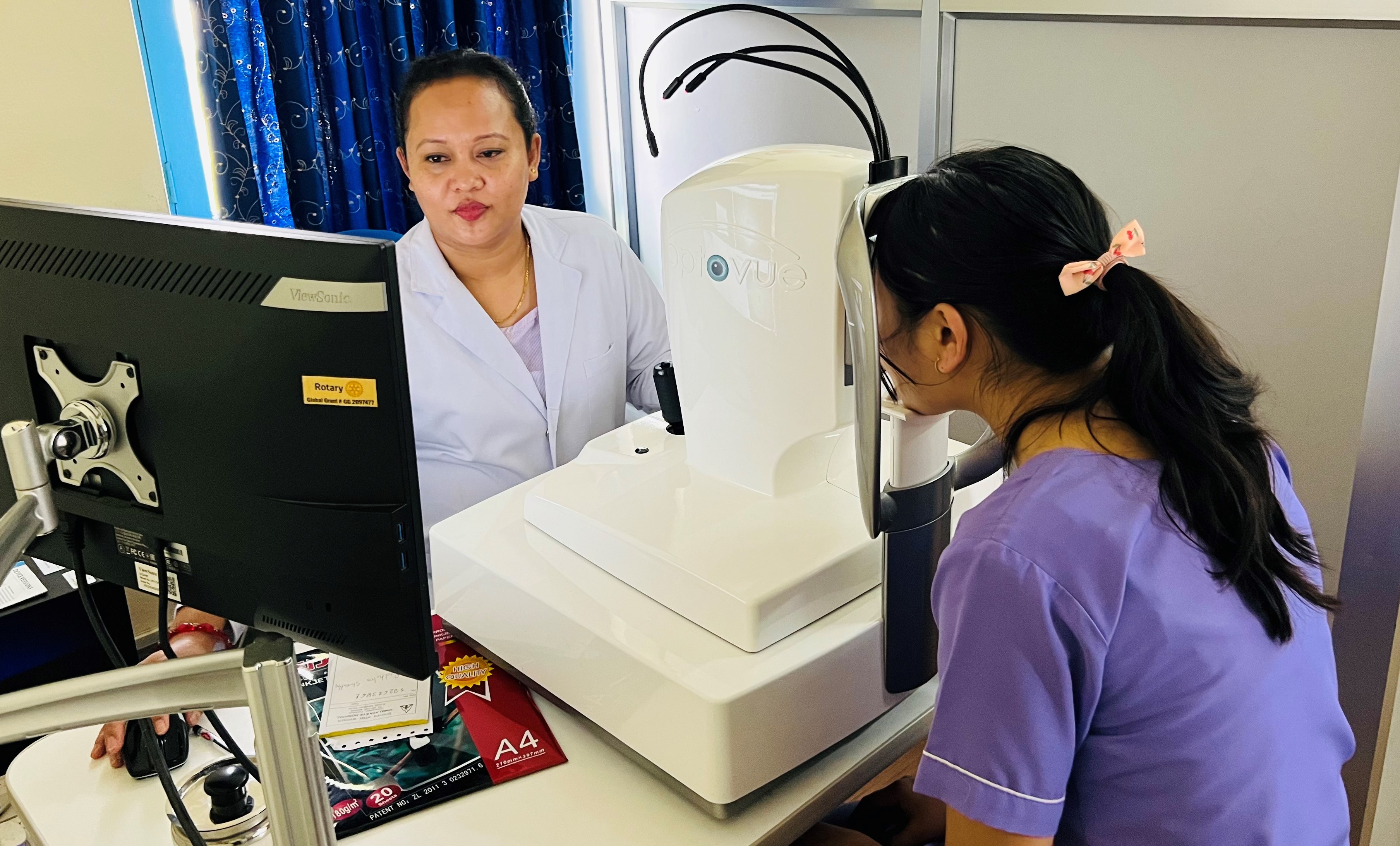
- Optical Coherance Tomography Angiography (OCT - A): We have latest and advance OCT-A including anterior OCT and noninvasive angiography retina, disc, non-invasive technique for imaging the micro vasculature of the retina and the choroid.
- Fundus cameras: We have multi-functional, non mydriatic retinal camera easy to use auto focus, auto shoot and auto small pupil detector options for quick and easy image acquisition - adds fluorescein angiography (FA) imaging.
- Humphrey field analyzer (HFA): The results of the analyzer identify the type of vision defect. Therefore, it provides information regarding the location of any disease processes or lesions throughout the visual pathway.
- Optical Biometer - We have latest and advance Optical biometry is a non-contact automated modality used for measuring optical biometric parameters. In 10 seconds, six values for cataract surgery are measured. It aid easy to calculate IOL power.
- Corneal topography: Corneal topography is a procedure used to monitor and measure changes that may occur to the shape and integrity of the cornea of your eye. This information provided in the testing illustrates corneal astigmatism, detection of corneal pathologies and perfection of contact lens fitting. Additional facility is to check Meibomian gland cells and noninvasive dry eye
- Vision electro physiology (ERG, VEP, EOG): Visual electro physiology is an extremely useful tool to study the retinal function objectively. It assesses the functional aspects of not only the retina but also the visual pathway as a whole. Even in this era of advanced imaging, one cannot ignore the importance of the functional aspects of the cellular network in the retina. They give us information about functional integrity of the visual system and assess the disorders affecting visual pathway, retina, optic nerve and higher visual processing center of the brain. Along with advanced imaging it gives an additional information of various ocular diseases. There are different type of electro physiological test which include electro retinography (ERG), electro oculography (EOG), and visual evoked potential (VEP). .